Cardiologists and surgeons may soon have a new cardiac imaging tool to improve the clinical outcomes of patients who need pacemakers, coronary artery bypass surgery, or angioplasty. The results of this study were published online in the Journal of the American College of Cardiology: Cardiovascular Imaging. A study conducted by Dr. James White of the University of Western Ontario and his colleagues invented a new imaging technology that can provide a single, 3D high-resolution heart image that clearly displays the vasculature and scar tissue in the myocardium. The study used a 3-Tesla MRI and was conducted at the Western Robards Institute. Heart attack or inflammation can cause permanent damage or scarring of the heart muscle. "We already know that MRI can image myocardial (heart) scar tissue, but this new technology has reached a new level of scar tissue imaging," Dr. White explained. "This is the first time we can see myocardial scars and heart blood vessels at the same time, and can build a three-dimensional model of the human heart, helping us quickly understand the relationship between heart blood vessels and related permanent damage. This will help guide surgeons and cardiology Experts better target blood vessels that respond to treatment, rather than blood vessels in irreversible muscle lesions. " This technique obtained the first three-dimensional coronary image using continuous contrast agent gadolinium perfusion, in which the blood pool is bright and clear. When the contrast agent is injected into the blood, 3-T MRI is used for imaging to provide high-resolution, three-dimensional images of coronary arteries. Although the contrast agent in the blood and normal tissues has been completely discharged, the signal of the scar tissue can be maintained because the contrast agent is slowly discharged from the scar tissue. After repeating the camera for 20 minutes, the scar tissue was highlighted and the image was three-dimensional. Since the two images of scar tissue and coronary vessels use the same method of MRI pulse sequence, they are completely suitable for fusing one of them to the other. A three-dimensional model of the heart showing blood vessels and scar tissue can now be created. The imaging technique was tested on 55 patients undergoing bypass surgery or cardiac pacemaker installation treatment called cardiac resynchronization therapy (CRT). The results of the study showed that the imaging technique is clinically feasible. This new imaging technique may have valuable value for patients planning blood vessel-based cardiac intervention. Dr. White described that the value of bypass surgery or angioplasty depends on whether to open the blocked blood vessel. If scar tissue can be seen in that area, there is no expected benefit. The scarred myocardial area reached by the CRT pacemaker leads will prevent the benefit of this treatment. The research fund was provided by the Ontario Heart and Stroke Foundation and the Canadian Innovation Foundation
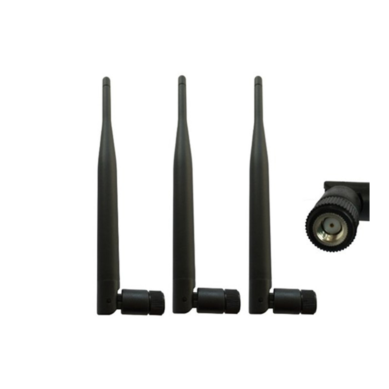
The Description of 3G Rubber Antenna:
The 3G Rubber Antenna is a multiband Covert Omni Antenna that allows the use of multiple frequency bands with a single antenna. Suitable for Frequencies: HSDPA, UMTS (3G) (1920-2770 MHz), GSM (2G) 900 (824-960 MHz), DCS 1800 (1710-1880 MHz)
3G Rubber Antenna Polarization: Vertical | 3G Rubber Antenna Connector Type: Customized | Impedance:50 ohm | VSWR:2 Input
The Advantage of 3G Rubber Antenna:
1,Rich experience , proven technique
2,Reasonable price
3,Wider frequency
4,OEM/ODM available
5,Perfession servie
6,ISO:9001 certification
7,1 year guarantee
8,Fast delivery time
9,R&D
10,Huge Capacity
3G Rubber Antenna 3G Rubber Antenna,Black Rubber Antenna,Folding Rubber Antenna,Omni 3G Antenna Shenzhen Yetnorson Technology Co., Ltd. , http://www.yetnorson.com